Scientists say they’ve identified a new type of cartilage — one that was actually discovered in the 19th century, forgotten, rediscovered and then forgotten again.
Medical textbooks describe three types of cartilage: hyaline, elastic and fibrocartilage. Hyaline cartilage helps bones slide smoothly over each other at the joints; elastic cartilage is very flexible and found in the external ear, voice box, and tube between the ear and throat; and fibrocartilage is tough and absorbs impacts at the joints and in the spine. The cells within these tissues are surrounded by lots of collagen and elastic fibers, the proportions of which give each type of cartilage its distinct characteristics.
Now, however, scientists say there’s a fourth type of cartilage that looks very different from the other kinds.
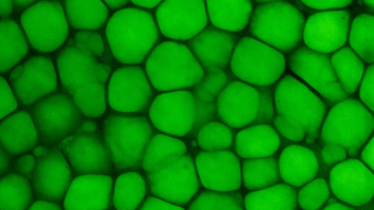
The tissue, which they call “lipocartilage,” superficially looks a lot like adipose tissue — better known as fat. It contains bulbous, balloon-like cells filled with oils, and the cells are surrounded by a thin matrix of fibers, rather than a thick matrix common to other cartilage. The cells are also highly uniform and can pack close together like bricks. Together, the cells form a springy, squishy tissue that has a bit of give but still resists deformation and tearing; this tissue is found in structures like the external ear and nose.
Related: New part of the body found hiding in the lungs
Some experts were impressed with the new analysis of lipocartilage. For instance, Viviana Hermosilla Aguayo and Dr. Licia Selleri of the University of California, San Francisco wrote in a commentary that this “long-ignored cartilage type” may “warrant updates to histology and anatomy textbooks.”
Others said that the researchers provided strong evidence of the tissue’s existence but that they aren’t sure lipocartilage merits its own classification.
“The authors provided evidence that this lipid-containing cartilage tissue exists in multiple mammals, including humans,” Shouan Zhu, director of the Osteoarthritis Research Laboratory at Ohio University, who was not involved in the study, told Live Science in an email. But “what I am not sure [of] is if it should be considered as a separate new type of tissue or just a new feature of an existing tissue,” namely, elastic cartilage.
What’s old is new again
“This was a serendipitous discovery,” study senior author Maksim Plikus, a professor in the Department of Developmental and Cell Biology at the University of California, Irvine, told Live Science in an email. The team was studying the skin of mouse ears when they stumbled upon the fat-stuffed cartilage cells, which Plikus compares to “Bubble Wrap.”
But when they dug deeper, the team learned their discovery wasn’t entirely novel. It turns out that other scientists had spotted this unique tissue before, Plikus noted.
In the 1850s, histologist Franz von Leydig wrote about his observations of rats’ ear cartilage under the microscope. “At first sight it looks like adipose tissue,” but it still has a distinct matrix, like cartilage, he noted. Leydig’s observations would fall into obscurity for more than a century. Then, in the 1960s, scattered reports described similar fatty tissues in rodent ears. In 1976, a pair of scientists coined the term “lipochondrocyte” for the cells found in lipocartilage. But again, these discoveries were soon forgotten.
Now, with their study published Thursday (Jan. 9) in the journal Science, Plikus and colleagues offer a very close analysis of lipocartilage, revealing its stages of development, genetic traits and molecular characteristics. The study highlights how the tissue differs from its look-alike — fat — and how it’s similar to other types of cartilage.
In mice, the team showed that the structures that hold the fat within lipocartilage are “superstable.” Unlike fat cells, which get larger or smaller depending on food intake, lipocartilage cells don’t shrink in times of starvation or swell in response to surplus. This stability partly stems from the tissue’s lack of enzymes for breaking down fats, as well as from a lack of transporters that bring fats from food into the tissue, the team found.
“This characteristic may offer an evolutionary advantage for the ear pinna” — the outer ear — “by enhancing its ability to gather and focus acoustic waves,” Zhu said. Sound waves ripple through fat very efficiently, so perhaps maintaining this high-fat cartilage in the external ear is useful for hearing, the authors also suggested.
Since the lipocartilage in the ear isn’t going to swell or shrink depending on calorie intake, the acoustics it supports should also stay steady over time.
Where is lipocartilage found?
The study authors initially identified lipocartilage in the external ears, noses and throats of mice. “It is in the nasal cartilage at the very tip of the nose,” Plikus said. “It forms the entire ear cartilage,” as well as the vast majority of larynx, or voice box, he said.
“In all these places, a high degree of elasticity is required and unlike the body’s other cartilages, such as joint cartilage, these structures are not weight-bearing,” he added. For example, a structure in the throat called the epiglottis flexibly flaps back and forth during swallowing to stop food from getting into the respiratory tract.
After studying mice, the researchers also identified lipocartilage in human fetal tissues, namely from the ear, nose, epiglottis and thyroid cartilage, which sits above the thyroid gland. They also found that lipocartilage emerged in models of human cartilage that they grew from stem cells in the lab.
To see how prevalent lipocartilage is across the animal kingdom, the team examined museum specimens of dozens of species. They spotted the tissue in several mammals — such as the Cairo spiny mouse (Acomys cahirinus), squirrel glider (Petaurus norfolcensis) and Pallas’s long-tongued bat (Glossophaga soricina) — but didn’t find it in any nonmammals, such as frogs, birds or alligators.
The authors say questions remain about when lipocartilage first emerged and what evolutionary advantages it might offer to the animals it’s found in. The team hopes to study the tissue’s evolution, as well as investigate its ability to regenerate after injury and probe whether lipocartilage contains different subtypes of cells. They also want to better understand how the cells manage such high fat content, “which can be toxic for many other cell types,” Plikus said.
“We believe these findings of a new cell type and new tissue type are fundamental and paradigm-shifting,” he said of the work so far.


