



The brain develops in stages, and it’s important that certain cells form before others so that precise neural connections can emerge later. But miniature models of the brain, called organoids, may not share this same sense of time, a new study finds.
These brain organoids are clusters of lab-grown neurons that mimic structural and functional aspects of full-size brains. Some scientists use these “minibrains” to study brain disorders that can arise during fetal development.
If the precisely timed processes involved in brain development go wrong, it can lead to conditions like macrocephaly and microcephaly in which the brain grows abnormally large or small, said study co-author Simon Hippenmeyer, a neuroscientist at the Institute of Science and Technology Austria.
Latest Videos FromLive Science
Because it’s not possible to directly study these conditions in human fetuses, scientists like Hippenmeyer turn to brain organoids. But the new study, published Aug. 12 in the journal Nature, suggests that organoids may be missing some crucial ingredients that would help them resemble real brains.
A matter of time
In earlier experiments in mice, Hippenmeyer’s team tracked the stem cells that make neurons, called radial glial progenitors (RGPs), during early embryonic development. They did this by giving pregnant mice a drug that activated fluorescent labels on individual stem cells. Then, they traced the resulting lineages of those stem cells after birth to see how many cells they produced and how that number changed as development progressed.
In the new study, they used that data as a reference to compare with brain organoids grown from mouse embryonic stem cells. They aimed to see if the developmental sequence in the organoids resembled that in the real brains.
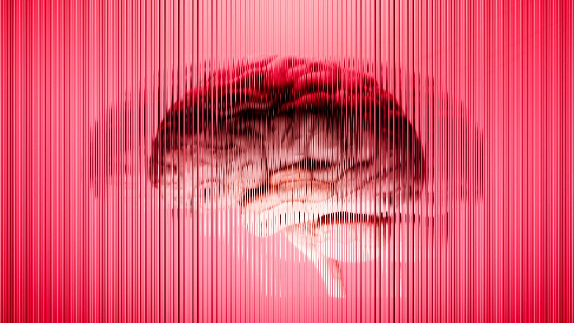
The researchers tracked how neurons developed in embryonic mouse brains and then compared those findings to what they observed in brain organoids.
(Image credit: JUAN GAERTNER/SCIENCE PHOTO LIBRARY via Getty Images)
The organoids produced the same major types of cells as the real brain did, the team found. However, the sequence of development was not preserved.
Get the world’s most fascinating discoveries delivered straight to your inbox.
During normal brain development, RGPs in the cerebral cortex — the brain’s outermost tissues — divide to increase their numbers and then differentiate, forming neurons and glial cells, which help to support, nourish and insulate neurons. In the organoids, however, some RGPs formed neurons too early, while others were still proliferating.
Normally, RGPs from later stages of development produce fewer descendants than the stem cells do in early development. But in the organoids, they produced more descendants regardless of the developmental time frame.
After seeing these patterns, the team investigated the variety of neurons that a single RGP could produce in an organoid. The cortex is made up of six layers of tissue, and normally, an individual RGP can produce both deeper-layer neurons (earlier in development) and upper-layer neurons (in the later stages). But in the organoids, about one-third of the RGPs became restricted to just one of these fates and then couldn’t make the other type of neuron.
In a real brain, Hippenmeyer noted, there are blood vessels, extracellular structures and metabolic signals that affect the organ’s development but are absent in the organoid. The researchers think these external signals may help coordinate the timing of neuronal development and the later formation of neural circuits.
“This suggests that there is something about the local environment of the cells [in the brain] that gives rise to the neurons,” said Denis Jabaudon, a neurobiologist at the University of Geneva who was not involved in the study. However, scientists don’t have a precise understanding of what those signals are yet, Jabaudon said.
For neurons to find each other, they have to be in the right place at the right time, Jabaudon added. “So, if you shift the timing of development, you’re going to be shifting the opportunities for connections and the opportunities for specific circuits” to form, he told Live Science.
Identifying what’s missing from these organoids will involve identifying and investigating those external signals neurons receive in the live brain. Then, scientists can identify differences between real brains and organoids that inform us about how the process naturally happens, Hippenmeyer said.
Once these missing ingredients are identified, this could “open the door” to systematically add back those signals in organoids in a way that would mimic the real brain closely, he told Live Science. Hippenmeyer added that the team also plans to grow brain organoids with human cells and investigate the same questions.
“We are very keen on finding out how those radial glial stem cells would behave in a human system,” he told Live Science.
Stouffer, M., Miranda, O. A., Pauler, F. M., Pipicelli, F., Streicher, C., Cheung, G., & Hippenmeyer, S. (2026). Temporal uncoupling of radial glia lineage progression in cortical organoids. Nature. https://doi.org/10.1038/s41586-026-10916-7
See how much you know about the most complex organ in the human body with our brain quiz!